


Consultation
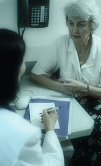 Your treating doctor will take a detailed history from you
to establish not only what is wrong (make a diagnosis),
but also determine the presence of any risk factors, especially
for Breast Cancer. They can do this in a caring and empathic
manner, putting the patient at ease and re-assuring them
that they will have the best of care and treatment if needed.
Your treating doctor will take a detailed history from you
to establish not only what is wrong (make a diagnosis),
but also determine the presence of any risk factors, especially
for Breast Cancer. They can do this in a caring and empathic
manner, putting the patient at ease and re-assuring them
that they will have the best of care and treatment if needed.
Consultations are chaperoned by our Breast Nurse but do not hesitate to bring your partner, spouse or friend with you, if that’s what you would like.
A Breast Clinic is not the ideal place for little children, so we ask that, where possible, other arrangements be made to care for them during your consultation.
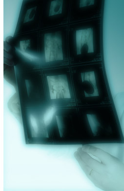
 This
is the investigation of the tissue of the breast by a
specialized and very sensitive X-ray technique and will
be done for you, if appropriate, immediately following
your consultation. The breast nurse will escort you down
to Benson’s Radiology where your mammogram (and
or ultrasound) are done and given to you to bring back
to Doctor.
This
is the investigation of the tissue of the breast by a
specialized and very sensitive X-ray technique and will
be done for you, if appropriate, immediately following
your consultation. The breast nurse will escort you down
to Benson’s Radiology where your mammogram (and
or ultrasound) are done and given to you to bring back
to Doctor.
The examination involves having the breast flattened between two plates allowing a detailed soft – tissue image of the structure of the breast. It can be momentarily uncomfortable but most patients find this very tolerable. It is the most proper investigation for Breast Cancer in women over 40 years of age. Other techniques, such as ultra-sound and MRI may or may not be recommended.
It is most important to bring any previous mammograms that your may have had with you to the consultation, thus allowing comparisons to be made with any new X-rays. Always bring your mammograms with you to clinic.
Ultrasound
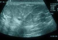 Ultra-sound
is the imaging of tissues by high frequency sound waves
and is most useful in the diagnosis and management of
Breast and Endocrine diseases. Diagnostic ultra-sound
will be done for you in Benson’s Radiology, unless
you have had one recently, often at the same time as
your mammogram. This is a painless procedure and does
not involve compression of the breast. It is the best
way to assess breast tissues for cysts, and is the investigation
of choice in the younger woman where mammography is not
so accurate.
Ultra-sound
is the imaging of tissues by high frequency sound waves
and is most useful in the diagnosis and management of
Breast and Endocrine diseases. Diagnostic ultra-sound
will be done for you in Benson’s Radiology, unless
you have had one recently, often at the same time as
your mammogram. This is a painless procedure and does
not involve compression of the breast. It is the best
way to assess breast tissues for cysts, and is the investigation
of choice in the younger woman where mammography is not
so accurate.
Doctor also has a “table – top” ultrasound in clinic to help visualize lesions in the breast or other tissues to allow aspiration, fine needle or core biopsy or ongoing measurement and assessment of lesions not removed.
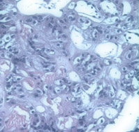
Needle Biopsy
Lesions visualized or palpable within the breast can
be sampled for Pathological assessment. Sampling
can be by “Fine Needle” aspiration, where
a sample is drawn from the area of concern into a
fine needle and then spread onto a slide for the
pathologist for assessment by cytology. This involves
a pin prick, not unlike having a blood sample taken,
and is well tolerated and usually not uncomfortable.
Doctor may do it using the table-top ultrasound to
be sure that the needle is placed into the lesion
accurately.
Sampling can also be by “Core Biopsy”. This involves the use of a larger size of needle and often requires a small dose of local anesthetic. Larger, and thus more diagnostically accurate, tissue samples are taken and given to the pathologist for assessment by histology which usually takes 24 hours.
 Accuracy
of Diagnosis
Accuracy
of Diagnosis
It is most important to recognize the accuracy of our diagnostic maneuvers is
not 100%. A “triple test” is done for all breast lesions and does
usually make a diagnosis, but it is not infallible. Doctor will always advise
a follow – up visit for re-assessment of your condition or lump.
It is essential to attend for that follow-up appointment, even if the initial diagnosis is benign, or the lump appears to have settled. Breast Cancer can occasionally be very difficult to diagnose and appear benign in its early stages. Failure to attend may result in a delay in diagnosis of a malignant condition and be detrimental to your health, outcome and survival.